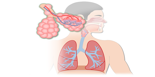
DIVISIONS OF THE RESPIRATORY SYSTEM
The respiratory system may be divided into the upper respiratory tract and the lower respiratory tract. The upper respiratory tract consists of the parts outside the chest cavity: the air passages of the nose, nasal cavities, pharynx, larynx, and upper trachea. The lower respiratory tract consists of the parts found within the chest cavity: the lower trachea and the lungs themselves, which include the bronchial tubes and alveoli. Also part of the respiratory system are the pleural membranes and the respiratory muscles that form the chest cavity: the diaphragm and intercostal muscles. Have you recognized some familiar organs and structures thus far? There will be more, because this chapter includes material from all of the previous chapters. Even though we are discussing the body system by system, the respiratory system is an excellent example of the interdependent functioning of all the body systems.
PHARYNX
The pharynx is a muscular tube posterior to the nasal and oral cavities and anterior to the cervical vertebrae. For descriptive purposes, the pharynx may be divided into three parts: the nasopharynx, oropharynx, and laryngopharynx. The uppermost portion is the nasopharynx, which is behind the nasal cavities. The soft palate is elevated during swallowing to block the nasopharynx and prevent food or saliva from going up rather than down. The uvula is the part of the soft palate you can see at the back of the throat. On the posterior wall of the nasopharynx is the adenoid or pharyngeal tonsil, a lymph nodule that contains macrophages. Opening into the nasopharynx are the two eustachian tubes, which extend to the middle ear cavities.
LARYNX
The larynx is often called the voice box, a name that indicates one of its functions, which is speaking. The other function of the larynx is to be an air passageway between the pharynx and the trachea. Air passages must be kept open at all times, and so the larynx is made of nine pieces of cartilage connected by ligaments. Cartilage is a firm yet flexible tissue that prevents collapse of the larynx. In comparison, the esophagus is a collapsed tube except when food is passing through it
TRACHEA AND BRONCHIAL TREE
The trachea is about 4 to 5 inches (10 to 13 cm) long and extends from the larynx to the primary bronchi. The wall of the trachea contains 16 to 20 C-shaped pieces of cartilage, which keep the trachea open. The gaps in these incomplete cartilage rings are posterior, to permit the expansion of the esophagus when food is swallowed. The mucosa of the trachea is ciliated epithelium with goblet cells. As in the larynx, the cilia sweep upward toward the pharynx.
The right and left primary bronchi are the branches of the trachea that enter the lungs. Their structure is just like that of the trachea, with C-shaped cartilages and ciliated epithelium. Within the lungs, each primary bronchus branches into secondary bronchi leading to the lobes of each lung (three right, two left). The further branching of the bronchial tubes is often called the bronchial tree. Imagine the trachea as the trunk of an upside-down tree with extensive branches that become smaller and smaller; these smaller branches are the bronchioles. No cartilage is present in the walls of the bronchioles; this becomes clinically important in asthma. The smallest bronchioles terminate in clusters of alveoli, the air sacs of the lungs.
Alveoli
The functional units of the lungs are the air sacs called alveoli. The flat alveolar type I cells that form most of the alveolar walls are simple squamous epithelium. In the spaces between clusters of alveoli is elastic connective tissue, which is important for exhalation. Within the alveoli are macrophages that phagocytize pathogens or other foreign material that may not have been swept out by the ciliated epithelium of the bronchial tree. There are millions of alveoli in each lung, and their total surface area is estimated to be 700 to 800 square feet. Each alveolus is surrounded by a network of pulmonary capillaries.
PULMONARY VOLUMES
The capacity of the lungs varies with the size and age of the person. Taller people have larger lungs than do shorter people. Also, as we get older our lung capacity diminishes as lungs lose their elasticity and the respiratory muscles become less efficient. For the following pulmonary volumes, the values given are those for healthy young adults.
SUMMARY
As you have learned, respiration is much more than the simple mechanical actions of breathing. Inhalation provides the body with the oxygen that is necessary for the production of ATP in the process of cell respiration. Exhalation removes the CO2 that is a product of cell respiration. Breathing also regulates the level of CO2 within the body, and this contributes to the maintenance of the acid–base balance of body fluids. Although the respiratory gases do not form structural components of the body, their contributions to the chemical level of organization are essential to the functioning of the body at every level.